Confocal and Super Resolution Microscopes
-
Nikon AXR-NSPARC Confocal and Super Resolution Microscope
Show More
-
The Nikon AXR-NSPARC is an inverted laser scanning confocal microscope that provides high-resolution fluorescence imaging and volume reconstructions. This microscope is also equipped with a Nikon Spatial Array Confocal (NSPARC) detector that allows super-resolution imaging of standard fluorescence samples down to 100 nm lateral resolution and 300 nm axial resolution.
Specifications:
- Objectives:
- Plan Apochromat λ D 2x; NA - 0.1; WD - 8.5 mm
- Plan Apochromat λ D 4x; NA - 0.2; WD - 20 mm
- Plan Apochromat λ D 10x; NA - 0.45; WD - 4.0 mm
- Plan Apochromat λ D 20x; NA - 0.8; WD - 0.8 mm
- Plan Apochromat λ S 40x (Silicone); NA - 1.25; WD - 0.3 mm
- Plan Apochromat λ D 60x (Water); NA - 0.127; WD - 0.18 mm
- Plan Apochromat λ D 60x (Oil); NA – 1.42; WD - 0.15 mm
- Lasers: 405 nm, 445 nm, 488 nm, 514 nm, 561 nm, 640 nm
- Tunable spectral emission detection for confocal imaging
- Filters for NSPARC emission detection: blue, green, red, far red, and a quad bandpass filter
- Resolution with NSPARC: 100 nm lateral and 300 nm axial
- High-speed resonant scanner
- Piezo motorized stage
- Focus control for long-term imaging
- Stage-top incubation
- Inverted microscope stand
- Compatible with glass slides, 35 mm dishes, 60 mm dishes, and multi-well plates
Applications:
- Confocal microscopy
- Live cell imaging
- Super-resolution microscopy
- Z-stacking
- Spectral unmixing
- Time-lapse imaging
- Differential Interference Contrast (DIC)
- Transmitted light imaging
- Organoid imaging
- Fluorescence recovery after photobleaching (FRAP)
- Fluorescence resonant energy transfer (FRET)
- Tiling of large image areas
- Fluorescence correlative spectroscopy
- BSL-2 samples
- Objectives:
-
Nikon AXR Upright Confocal Microscope
Show More
-
This Nikon AXR is an upright laser scanning confocal microscope that provides high-resolution fluorescence imaging and volume reconstructions. Notably, this microscope has a water dipping objective, which allows imaging with the front lens immersed in the sample media.
Specifications:
- Objectives:
- Plan Apochromat λ D 4x; NA - 0.2; WD - 20 mm
- Plan Apochromat λ D 10x; NA - 0.45; WD - 4.0 mm
- Plan Apochromat λ D 20x; NA - 0.8; WD - 0.8 mm
- Plan Fluor 40x (Oil); NA - 1.3; WD - 0.2 mm
- Plan Apochromat λ D 60x (Oil); NA – 1.42; WD - 0.15 mm
- Plan Fluor λ D 10x (water dipping); NA - 0.3; WD - 3.5 mm
- Lasers: 405 nm, 445 nm, 488 nm, 514 nm, 561 nm, 640 nm
- Tunable spectral emission detection
- High-speed resonant scanner
- Upright microscope stand
- Compatible with glass slides, 35 mm dishes, and 60 mm dishes
Applications:
- Confocal microscopy
- Water dipping objective
- Z-stacking
- Spectral unmixing
- Time-lapse imaging
- Differential Interference Contrast (DIC)
- Transmitted light imaging
- Fluorescence recovery after photobleaching (FRAP)
- Fluorescence resonant energy transfer (FRET)
- Tiling of large image areas
- Cleared tissue imaging
- Objectives:
-
Nikon A1R Confocal Microscope
Show More
-
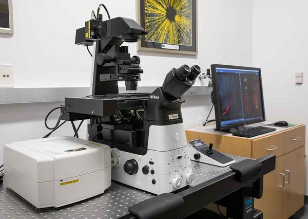
The Nikon A1R confocal microscope provides high-resolution fluorescence imaging using a variable pinhole for optical Z sectioning and either galvano or high-speed resonant scanning (30-420 fps). The system is equipped with the following excitation wavelengths: 405, 445, 488, 514, 561, and 640 nm, as well as filter sets for DAPI, CFP, GFP, YFP, TRITC, and Cy5. It also has ultra-sensitive GaAsP and high sensitivity PMT detectors and Perfect Focus control, which maintains the focal plane over long imaging experiments.
Objectives:
- Plan Apo λ 10x; NA - 0.45; WD - 4 mm
- Plan Apo λ 20x; NA - 0.75; WD - 1 mm
- Plan Fluor DIC 40x (Oil); NA - 1.3; WD - 0.24 mm
- Plan Apo λ 60x (Oil); NA - 1.4; WD - 0.14 mm
Incubation: An incubation stage for temperature and CO2 control for long-term live cell imaging experiments is available.
Location: This microscope is located in HDB 3.222KB.
Instrument Access: Assisted use and training for individual use are available. To schedule assisted use or training, please email Anna Webb.
Fees: trained users: $53/hour; assisted use: $123/hour
-
Nikon W1 Spinning Disk Confocal Microscope
Show More
-
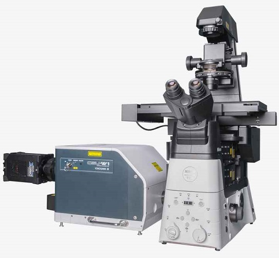
The Nikon W1 spinning disk confocal microscope uses a Yokogawa W1 scanning head with multiple pinholes for ultrafast confocal imaging. The W1 has an extra wide field of view, making it ideal for imaging large specimens. It has the following excitation lasers: 405, 488, 561, and 640 nm, and filters for DAPI, GFP, Texas Red, and Cy5. It is equipped with dual monochromatic Andor EMCCD cameras for simultaneous two-color imaging. It also has Perfect Focus control and DIC optics.
Objectives:
- Plan Fluor DLL Ph1 10x; NA - 0.3; WD - 16.0 mm
- Super Plan Fluor Ph1 ELWD 20x; NA - 0.45; WD - 6.9-8.2 mm
- Plan Apochromat λ 20x; NA - 0.75; WD - 1.0 mm
- Super Plan Fluor ELWD 40x; NA - 0.6; WD - 2.8-3.6 mm
- Plan Apochromat VC 60x (Water); NA - 1.20; WD - 0.28-0.31 mm
- Plan Apochromat λ 100x (Oil); NA - 1.45; WD - 0.13 mm
Incubation: An incubation stage for temperature and CO2 control for long-term live cell imaging experiments is available.
Location: This microscope is located in MBB 1.426T.
Instrument Access: Assisted use and training for individual use are available. To schedule assisted use or training, please email Anna Webb.
Fees: trained users: $53/hour; assisted use: $123/hour
-
Nikon TIRF/N-STORM System
Show More
-
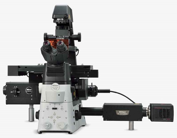
The TIRF/N-STORM System has a single inverted microscope stand that functions as both a TIRF microscope and super resolution N-STORM system.
The shared Nikon Eclipse Ti-2 microscope stand includes:
- Objectives:
- Plan Apochromat λ 10x; NA - 0.45; WD - 4.0 mm
- Plan Apochromat λ 20x; NA - 0.75; WD - 1.0 mm
- Plan Fluor 40x (Oil); NA - 1.3; WD - 0.20 mm
- Plan Apochromat λ 60x (Oil); NA - 1.4; WD - 0.13 mm
- High Power TIRF Auto Correction Collar 100x (Oil); NA - 1.49; WD - 0.12 mm
- Incubation: The microscope has incubation controls for both temperature and CO2 for live cell imaging.
- Focus control: The microscope is equipped with Perfect Focus to automatically correct focus drift.
The Total Internal Reflectance Fluorescence (TIRF) microscope provides high signal-to-noise fluorescence imaging of a thin plane at the cell-coverslip interface. This microscope is equipped with filters for DAPI, GFP, Texas Red, and Cy5. It has two monochromatic EMCCD cameras that can be used for single color imaging or simultaneous two color imaging. It is equipped with the following laser excitation wavelengths: 405, 488, 561, and 640 nm.
The N-STORM (Stochastic Optical Reconstruction Microscopy) system uses a super resolution technique that images individual, blinking fluorophores over time and pinpoints the precise location of each fluorophore to reconstruct the image. It can achieve a lateral resolution limit of ~20 nm and axial resolution of ~50 nm. Multicolor imaging and 3D localization are possible with this microscope. This microscope is equipped with filters for DAPI, GFP, Texas Red, and Cy5. It uses a Hamamatsu sCMOS camera for high sensitivity detection of fluorophores. It is also equipped with the following laser excitation wavelengths: 405, 488, 561, and 640 nm.
Location: This microscope is located in MBB 1.426T.
Instrument Access: Assisted use and training for individual use are available. To schedule assisted use or training, please email Anna Webb.
Fees: trained users: $53/hour; assisted use: $123/hour
- Objectives: